
Biological Activities of Four Synthetized Peptides
- Open Access
- Peer Reviewed
- Similarity Checked
- CC BY 4.0
Abstract
Objectives
Bacterial resistance to conventional antibiotics is a serious public health problem. Over the past decade, small proteins known as antimicrobial peptides (AMPs), natural compounds produced by all prokaryotic and eukaryotic cells, have shown promising results in overcoming the growing problems of antibiotic resistance. There are nowadays several peptides of plant origin and rich in cysteines which have shown very good antimicrobial activities. The research of molecules with bactericidal activity is always current event with increase of multi resistance. The present study was undertaken to investigate the in vitro antibacterial activities of four synthesized peptides obtained in 97% yield by the solid phase synthesis method.
Method
The synthesis reaction was monitored by combined HPLC and LCMS methods and evaluation of the antibacterial property of these synthetized peptides. The antibacterial test was performed using micro dilution method on many species of bacteria, S. epidermidis ATCC 35984, S. aureus ATCC 25923, S. aureus ATCC 8095, E. faecalis ATCC 29212, E. faecium ATCC 700221, K. pneumoniae ATCC 700603, E. coli ATCC 25922, A. baumannii ATCC 19606, P. aeruginosa ATCC 27853.
Results
The conformity and effectiveness of the synthesis of the peptides was carried out by LCMS with an average yield of 91%. Peptides were bactericidal on almost all of these strains except E. coli ATCC 25922 where they were bacteriostatic. The biofilm eradication capacity test was carried out on two ATCC strains of S. epidermidis. Strain 35984 is a good biofilm-forming strain, while strain 12228 is considered a poor biofilm former. Comparison of the presence of biofilm between them showed a significant difference with p<0.05, showing that the controls used are significantly different in all cases.
Article Information
- Received
- Accepted
- Published
Academic Editor: Fernando Albericio, Research Professor at the School of Chemistry of UKZN.
Checked for plagiarism: Yes
Review by: Single-blind
Copyright © 2024 Olivier Ndogo Eteme, et al.
 This is an open-access article distributed under the terms of the Creative Commons Attribution License, which permits unrestricted use, distribution, and reproduction in any medium, provided the original author and source are credited.
Corresponding author: Olivier Ndogo Eteme, São Paulo State University (UNESP), School of Sciences and Engineering, Tupã, São Paulo, Brazil —
Competing Interests
Authors declare not conflict of interest.
Funding
No specific funding statement was provided by the authors.
Data Availability
No data-availability statement was provided by the authors.
Acknowledgements
This work is supported by São Paulo Research Foundation/FAPESP (Process number 2021/06706-9), FAPESP Post-Doctoral fellowship (Process number 2022/02808-4) and by the research group “Peptides: Synthesis, Optimization and Applied Studies - PeSEAp”.
Citation:
Introduction
Antibiotics are an effective treatment for a wide variety of infections and diseases 1. Unfortunately, the uncontrolled use of antibiotics in human medicine, agriculture and animal husbandry is the cause of the development of much resistance, resulting in a decreased ability to treat infections and diseases in humans, animals and plants 2, 3, 4. The appearance of these resistances to antibiotics causes crucial problems for the world population today 5; there is growing evidence that treatable illnesses, such as pneumonia, tuberculosis or minor infections, are becoming increasingly incurable in some patients, placing a greater economic and emotional burden on families and various healthcare systems, with increased human disease, suffering and death, increased cost and duration of treatment, and increased side effects associated with the use of multiple and more potent drugs 6, 7, 8. Ciprofloxacin, an antibiotic commonly used to treat urinary tract infections, has a resistance rate ranging from 8.4% to 92.9% for Escherichia coli and from 4.1% to 79.4% for Klebsiella pneumoniae9, 10, 11, 12, 13, 14. Another antibiotic, colistin, has had bacterial resistance detected in several countries and regions, causing infections for which there is currently no effective antibiotic treatment 15, 16. There is a very high rate of people with methicillin-resistant Staphylococcus aureus infections, of whom 64% are more likely to die than people with drug-sensitive infections 17, 18, 19. In the absence of effective methods to prevent and adequately treat drug-resistant infections, and without better access to existing and new antimicrobials of guaranteed quality, the number of deaths from treatment failures will continue to increase. Medical procedures, such as surgery, including cesarean sections or hip replacements, cancer chemotherapy and organ transplants, will become riskier. 20, 21, 22, 23. It is therefore important to develop new, more effective antibiotics, with reduced adverse effects, against the various multiresistant pathogens. New drug discovery essentially involves the identification of new chemical entities that exhibit the required characteristics of pharmacobility and medicinal chemistry 24. They can be generated either by chemical synthesis or by isolation from selected natural products. Antimicrobial peptides have been isolated and characterized from tissues and organisms representing virtually every kingdom and phylum, ranging from prokaryotes to humans 25. They can be mainly used for the rational design of bioactive molecules. They can act as ligands in the development of targeted treatments as well as diagnostics, can be used in vaccine design or can be used in agriculture 26. Several widely studied AMPs stand out as peptidase inhibitors. Proteolytic degradation of peptide-based drugs is often considered a weak point limiting systemic therapeutic applications. Therefore, enormous efforts are usually devoted to stabilizing sequences against proteases present in serum or plasma 27, 28. Many AMPs can inhibit various metabolic activities by inhibiting protease activity 29. For example, histatin 5 has a strong inhibitory effect on proteases secreted by the host and bacteria. The AMPs eNAP-2 and indolicidin inhibit microbial serine proteases, elastase and chymotrypsin 30. Cathelicidin-BF is a peptide isolated from the venom of Bungarus fasciatus, it can effectively inhibit thrombin-induced platelet aggregation and further block protease-activated receptor 4 31. Today, the scientific community increasingly recommends evaluating potential immunogenicity during the drug development process using diverse and varied approaches to manage the clinical consequences of immunogenicity 32. It is nevertheless important to mention that peptide drugs and their impurities can cause unexpected immunogenicity; the assessment of immunogenicity risks is always essential before marketing 33. It is therefore important to continue the search for new AMPs whose hydrolysis by proteases is reduced as much as possible.
Materials and methods
Antimicrobial Peptides Synthesis
The peptide synthesis was carried out following a method already described in the literature 34. Rink resin was used as a solid support for the synthesis. It was initially soaked in DMF for 30 minutes for activation. After that, it was then soaked in DCM for 30 minutes and then rinsed again in DMF. Deprotection of the fmoc group was done using a piperazine solution (10% piperazine w/v in DMF:EtOH 9:1). Deprotection consisted of introducing the piperazine solution into the resin, leaving it stirring for 1 minute, removing the solvent and adding new piperazine to the resin for 20 minutes then removing the solvent. Subsequently it was rinsed with DCM and DMF. The initial Fmoc deprotection step was carried out using a solution of piperazine in DMF. The different amino acids previously deprotected with piperazine as described previously were coupled to the resin for 2 hours at room temperature. The ninhydrin test made it possible to qualitatively control the effectiveness of the couplings. Cleavage of the synthesized peptides from the resin was done by hydrolysis using a solvent system containing TFA, TIS, EDT and water (94:1:2.5:2.5 v/v) for 2 hours at RT. Then, the samples were freeze-dried (Liotop, model K108, Brazil) until a powder was formed Scheme 1.
Scheme 1. Chemical mechanism synthesis of peptides catalysed by HoBt and DIC. 1. DIC activates the carbonyl of the carboxylic acid function of the coupled amino acid. The consequence is the reduction of a large number of side reactions. 2. After obtaining an activated intermediate, HOBt is used to produce activated esters. These esters are insoluble (like N-hydroxysuccinimide esters) and react with amines at room temperature to give amides 35.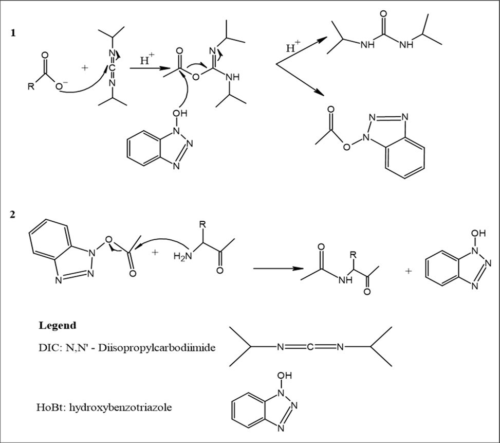
Download figure
Peptide Purification and Characterization
Analytical analysis was carried out following a method already described in the literature 34. Once the cleavage was completed, the resulting solution containing the synthesized peptide was lyophilized. A small quantity of the powder obtained was dissolved in water at a concentration of 5 mg/mL and was subjected to HPLC analysis (Simadzu Prominence, with a DGU-20A5R membrane degasser, an SPD-20A UV detector, a CTO-20A column oven, a SIL-10AF autosampler, an FRC-10A fraction collector and a qualitative LC-20AT double pump) for the purpose of verifying purity. The impure peptides were purified by injecting the samples through a C18 column with a flow of 5mL/min. Buffers A and B, respectively 0.045% TFA in aqueous solution (eluent A) and 0.036% TFA in acetonitrile (eluent B), were used as elution solvent. Verification of obtaining the desired peptide was done by LCMS and carried out on a Shimadzu chromatograph/Bruker spectrometer (Prominence/Amazon SL) with the same parameters as those used for the HPLC purification system at a flow rate of 0.5 mLmin−1.
Microbiological tests
Determination of Minimum Inhibitory Concentration – compounds soluble in DMSO
To determine the MIC, all strains of the bacterial species that were sensitive to the compound were used. Each compound was diluted in DMSO and a 100x concentrated stock solution was prepared; subsequently the stock solution was diluted 1:100 in Mueller Hinton Cation Adjusted (MHCA) broth (BD) according to CLSI 36. From this, each compound was tested in 512 μg/ml 1% DMSO, or at the highest concentration at which it was possible to dissolve the compound without precipitation. From the compound wells at 512 μg/ml, serial dilutions (1:2) were made up to a concentration of 0.06 μg/ml. The incubation was carried out at 37°C and the results were visually read after 24 hours, in which it was observed up to which concentration the compound was able to inhibit the growth of the microorganism. For negative and positive controls, Mueller Hinton Cation Adjusted broth 1% DMSO was added. In the positive control, bacteria without the compound were added to observe their growth in Mueller Hinton Cation Adjusted broth 1% DMSO. In the negative control, there is only the Mueller Hinton Cation Adjusted 1% DMSO broth culture medium, without bacteria, to show that there is no contamination thereof. Tests were performed in triplicate.
Determination of Minimum Inhibitory Concentration – water-soluble compounds
To determine the MIC, all strains of the bacterial species that were sensitive to the compound were used. Each compound was diluted in water and a 10x concentrated stock solution was prepared. Subsequently, the stock solution was diluted 1:10 in Mueller Hinton Cation Adjusted (MHCA) broth (BD), according to CLSI 36. From this, each compound was tested at 512 μg/ml or the highest concentration at which it was possible to dissolve the compound without precipitation. From the compound wells at 512 μg/ml, serial dilutions (1:2) were made up to a concentration of 0.06 μg/ml. The incubation was carried out at 37°C and the results were visually read after 24 hours, in which it was observed up to which concentration the compound was able to inhibit the growth of the microorganism. For negative and positive controls, Mueller Hinton Cation Adjusted broth was added. In the positive control, bacteria without the compound were added to observe their growth in Mueller Hinton Cation Adjusted broth. In the negative control, there is only the Mueller Hinton Cation Adjusted broth culture medium, without bacteria, to show that there is no contamination of it. Tests were performed in triplicate.
Determination of Minimum Inhibitory Concentration - Omnilog
To determine the MIC, all strains of the bacterial species that were sensitive to the compound were used. Each compound was diluted in DMSO and a 100x concentrated stock solution was prepared. Subsequently, the stock solution was diluted 1:100 in Mueller Hinton Cation Adjusted (MHCA) broth (BD), containing H dye (Biolog) in a proportion of 1.05%; the entire procedure was carried out following the recommendations of CLSI 36. From this, each compound was tested at 512 μg/ml, or at the highest concentration at which it was possible to dissolve the compound without precipitation. From the compound wells at 512 μg/ml, serial dilutions (1:2) were made up to a concentration of 0.06 μg/ml. Incubation was carried out with the aid of the OmniLog® device (Biolog, Hayward, CA, USA) at 36 °C ± 1 °C for 24h, followed by visual reading of the plates in which it was observed up to which concentration the compound was able to inhibit the growth of the microorganism. In the positive control, bacteria without the compound were added to observe their growth in Mueller Hinton Adjusted Cation broth – 1% DMSO. In the negative control, there is only the Mueller Hinton Cation Adjusted broth culture medium – 1% DMSO, without bacteria, to show that there is no contamination thereof; both controls also received the H dye in the same proportion. Tests were performed in triplicate.
Determination of Minimum Bactericidal Concentration
After visually reading the MIC, 100 μL of the contents of the well equivalent to the MIC, one dilution below and all dilutions above, were inoculated onto an MHCA-Agar plate using the microdrop technique, without streaking. The plate was incubated in an oven at 37°C for 24 hours, when a visual reading was made to observe which concentration did not result in bacterial growth. To determine bactericidal or bacteriostatic activity, compounds that had an MBC/MIC ratio less than or equal to four were considered bactericidal, above that being considered bacteriostatic 37.
Determination of biofilm eradication capacity
S. epidermidis ATCC 12228 (Negative control, poor biofilm former) and S. epidermidis ATCC 35984 (positive control, good biofilm former) were inoculated in BHI Broth 0.75% glucose in several replicates in a 96-well microplate. Incubation was carried out at 37°C for 24 hours to form the biofilm. The ability to eradicate the biofilm formed will be further tested after the formation of the S. epidermidis ATCC 35984 biofilm.
After 24 hours at 37ºC, the culture medium with the planktonic bacteria was removed and washed with 0.85% physiological solution so that only the biofilm remains on the microplate. Each compound was added at the highest possible concentration, diluted in 1% DMSO when necessary, in fresh BHI 0.75% glucose medium in the wells where biofilm was formed by S. epidermidis ATCC 35984. As a control for biofilm growth, 6 wells with the positive control and 6 negative controls should only receive BHI broth 0.75% glucose, plus 1% DMSO when necessary. Incubated at 37ºC for another 24 hours. This second step was to allow the biofilm to be eradicated by the compound.
After 24 hours, to quantify the biofilm, the wells were washed several times and stained with crystal violet. After washing steps and removal of excess violet crystal, the violet crystal was removed with ethanol:acetone (80:20) and transferred to another microplate for indirect quantification of the biofilm at 595 nm. The assay control was based on the comparison of the average absorbance of the S. epidermidis ATCC 35984 biofilm and the average absorbance of the S. epidermidis ATCC 12228 biofilm, both without compound. The comparison of means was carried out using the Student's T-Test, whose P value <0.05 proves the significant difference between two means from two different samples. Regarding the ability of the compound to eradicate biofilm, data analysis was based on the comparison of biofilm production of S. epidermidis ATCC 35984 without compound with S. epidermidis ATCC 35984 with compound. For this, the average of the replicates was used and the standard deviation was taken to analyze the coherence between the replicates. For statistical analysis, analysis of variance (ANOVA) was performed on the mean absorbance of biofilm production by the strain S. epidermidis ATCC 35984 without compound and the mean absorbance of biofilm production by S. epidermidis ATCC 35984 with each compound. When the analysis of variance had a value of P < 0.05, it indicated a significant difference between the two samples 38.
Hemolytic activity assay
Stock solutions of Alca1 were prepared, containing 1 mg resuspended in 0.5 mL of PBS buffer (pH 7.4). Serial dilutions of the peptides were performed, starting from a concentration of 512 µg /mL up to 1 µg/mL, all in duplicates. In addition, controls were prepared, with the negative control being PBS buffer and the positive control being 1% Triton. In addition, the erythrocyte solution was prepared by diluting it in PBS buffer (1/25). 100 µL of the erythrocyte solution was added to the tubes containing the peptide dilutions and incubated at 37ºC for 1 hour. Then, the tubes were centrifuged at 500 g for 5 minutes. The supernatants were pipetted into microplates for subsequent reading at 540 nm in a microplate reader.
To calculate the % hemolysis, the following were considered:

Proteolysis by pepsin, trypsin, chymotrypsin and endogluC
Pepsin, trypsin and chymotrypsin were dissolved in 10 mM phosphate buffer saline (PBS) (10 mM) by serial 10-fold dilution. The final concentrations of these two enzymes ranged from 2 × 10⁻⁶ mg/ml to 2 mg/ml. Peptides (at the concentration of 128, 256, and 512 μM, respectively) were mixed with different concentrations of trypsin or chymotrypsin. The samples were incubated for 6 h at 37°C. Then they were heated for 15 min at 60°C to terminate the enzyme reaction. We followed reaction with HPLC, every 30 min 39 by calculating, using the calibration curve, the concentration of each peptide remaining undegraded at a given time t. For endogluC, peptides were dissolved at 128, 256, and 512, 1000 μM in hydrolysis buffer (ammonium bicarbonate 100 mM, pH 7.8) and enzymatic hydrolysis was carried out for 16 h at 37 °C with an enzyme:substrate (E/S) ratio of 1:100 40. We followed the reaction with HPLC, every 30 min by calculating, using the calibration curve, the concentration of each peptide remaining undegraded at a given time t. The CD spectra were measured at 25 °C.
Prediction of potential antimicrobial regions was performed by obtaining a bactericidal propensity (PV) index value calculated for each amino acid in the AMPA software 41. The 3D structure of the peptides was modeled with the PEP-FOLD software. HeliQuest made it possible to calculate from the amino acid sequence of a helix (α-helix, 3-10 helix, 3-11 helix or π-helix) its physicochemical properties and its amino acid composition in order to identify the protein segments with similar characteristics 42.
Results
In this work, four peptides were proposed with antimicrobial properties. The physicochemical properties are represented below. The different antimicrobial peptides Neo1, Neo2, Neo3 and Neo4 were successfully obtained, with a purity greater than 97%. The three-dimensional structures of the alpha helices have been represented by two-dimensional projections that we call helical wheels. The revealed plot shows the hydrophobic amino acids concentrated on one side of the helix, and the polar or hydrophilic amino acids on the other. The different sequences of these peptides are shown below and the Schiffer-Edmundson projection in Figure 1A and the results of circular dichroism in Figure 1B. Monitoring of the effectiveness and purity of the synthesized peptides was obtained by HPLC and LCMS (Figure 1C).
Figure 1. A. Schiffer-Edmundson projection of different Neo peptides, B. CD spectra of peptides at a concentration of 80 µmol L−1 in 0.8 mmol L−1 of LUVs, C. Chromatogram HPLC et LCMS of Neo's peptides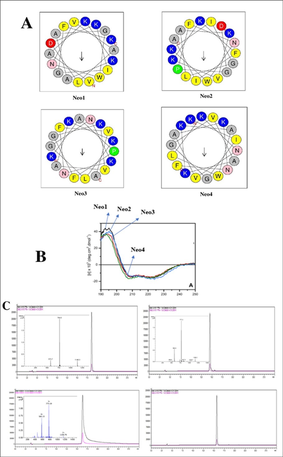
Download figure
Knowledge of certain physicochemical properties of peptides generally requires knowledge of their three-dimensional structure. It makes it possible to predict reaction sites, reactivity and complexation centers. Several software programs have already been developed, including PEP-FOLD. The algorithms designed in this software aim to use the letters SA of the structural alphabet to describe the conformations of four consecutive residues, coupling the predicted series of letters SA. The results of Neo's peptide analysis are shown below (Figure 2). A bactericidal propensity index was calculated for each amino acid, using as reference the experimental data reported by a high-throughput screening assay (Figure 3).
Figure 2. Pep-fold analysis of Neo's peptides. PEP-FOLD3 on-line interactive visualization of the models generated is based on the PV javascript protein viewer. Different representations as well as colouring schemes can be selected. A menu makes it possible to select a model among the 10 best models (representatives of the 10 best clusters). It corresponds to a graphical representation of the probabilities of each Structural Alphabet (SA). Letter (vertical axis) at each position of the sequence (horizontal axis). Note that SA letters correspond to fragments of 4 residue length. The profile is presented using the following color code: red: helical, green: extended, blue: coil.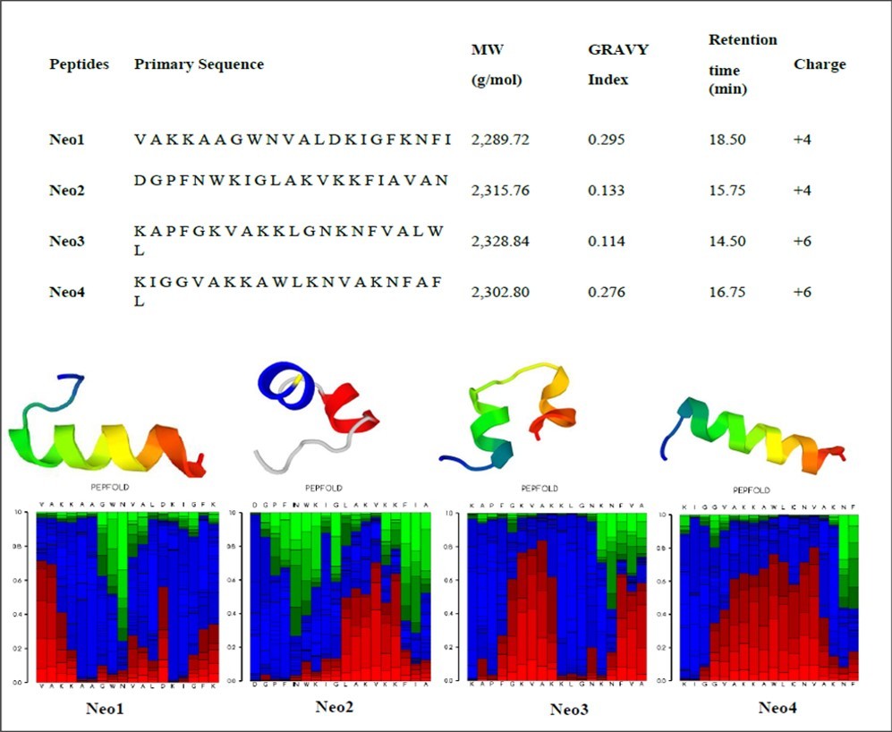
Download figure
Figure 3. Antimicrobial prediction of Neo's peptides. The above chart shows the antimicrobial profile for the entered sequence(s). On the X-axis is reported the amino acid position in the protein and on the Y-axis the antimicrobial score at that position compared to the other two proteases.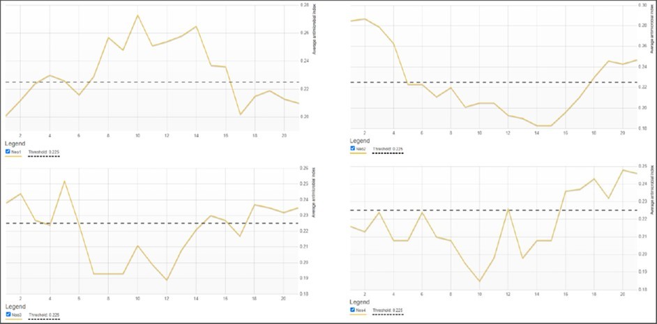
Download figure
The results of the enzymatic protease stability analyzes are summarized in Figure 4 below. From the analysis of these figures, the different peptides are relatively stable beyond one hour in contact with the different proteases pepsin, trypsin, chymotrypsin and endoglu-c. However, the Neo1 and Neo4 peptides present the same stabilities. They begin to degrade from the sixth hour with pepsin, chymotrypsin and endogluc and degrade from one hour on contact with trypsin.
Figure 4. Monitoring the kinetics of degradation of Neo1 to Neo4 peptides by pepsin, trypsin, chymotrypsin and endogluc. (1-4) consecutively represent the degradation digraph of Neo1 by pepsin, trypsin, chymotrypsin and endogluc. (5-8) consecutively represent the digraph of degradation of Neo2 by pepsin, trypsin, chymotrypsin and endogluc. (9-12) consecutively represent the degradation digraph of Neo3 by pepsin, trypsin, chymotrypsin and endogluc. (13-16) consecutively represent the degradation digraph of Neo4 by pepsin, trypsin, chymotrypsin and endogluc.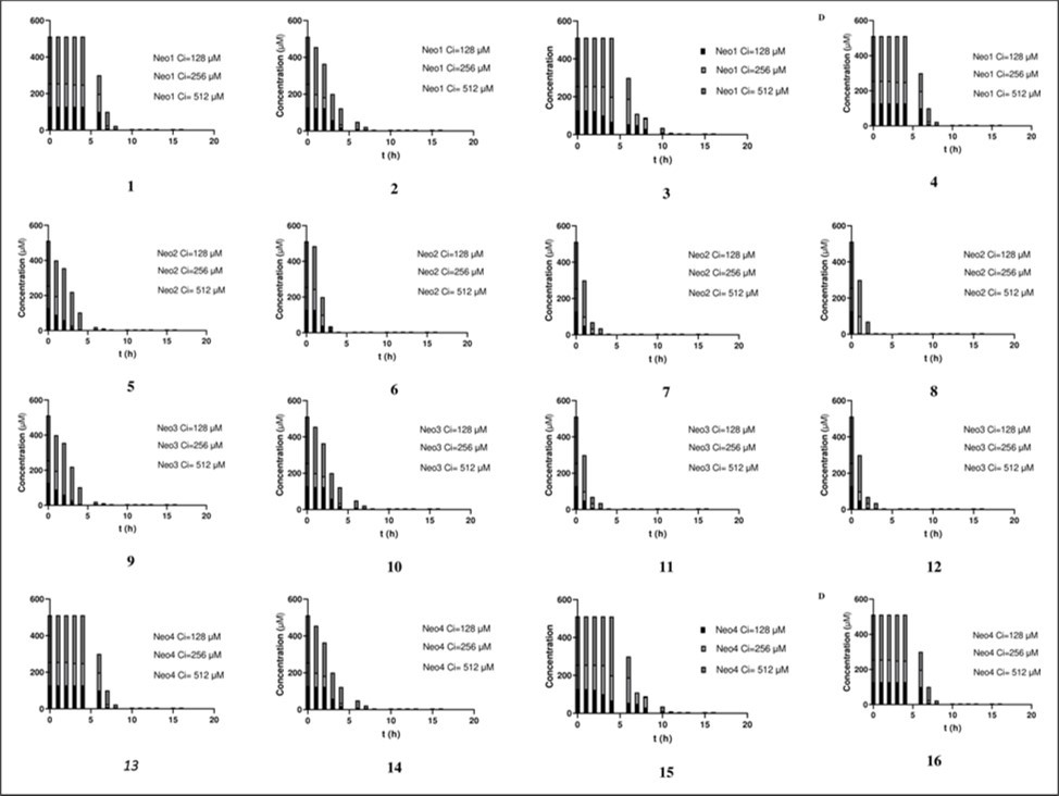
Download figure
The Neo2 peptide is less stable than the Neo1 and Neo4 peptides; it degrades after an hour with all the different proteases and is a little less sensitive to pepsin compared to the others.
The Neo3 peptide, like the Neo2 peptide, is less stable in contact with different proteases because it fades from the first hour; however, it is more stable to pepsin and trypsin.
Antibacterial activities of peptides against bacteria
Antimicrobial activity is defined by the CMB/MIC ratio. For ratios greater than 4, the activity is considered bacteriostatic, for results equal to or less than 4, the activity is considered bactericidal, according to Pankey and Sabath (2004). Results indicated with N.D., in the table 1 above, indicate that the activity cannot be defined and, N.R., indicates that the assay was not carried out with that strain as the compound showed no activity or presented an MIC value above 512 mg/L. Peptides Neo1 and Neo3 showed similar results as did Neo2 and Neo4.
To carry out a biofilm assay, two ATCC strains of S. epidermidis were used. Strain 35984 is a good biofilm-forming strain, while strain 12228 is considered a poor biofilm-former. The comparison of the presence of biofilm between them showed a significant difference with p<0.05, showing that the controls used are significantly different in all cases. The ability to eradicate the biofilm formed by the S. epidermidis ATCC 35984 strain was analyzed in the presence of each peptide. Some showed eradication results (p-value below 0.05), however, not showed eradication results above 50%. The other one exhibited a p-value lower than 0.05, however, it is important to highlight that this result does not suggest the eradication of the biofilm formed, but rather that it promoted such significant growth of the biofilm that it differentiated statistically from the control group, result is presented in the Table 1.
Table 1. MIC and MBC results for synthetized peptides| Bacterials strains | MIC (mg/L) | MBC (mg/L) | MBC/MIC | Activity* | ||||
|---|---|---|---|---|---|---|---|---|
| Neo1 | Neo2 | Neo1 | Neo2 | Neo1 | Neo2 | Neo1 | Neo2 | |
| S. epidermidis ATCC 35984 | 128 | 256 | 256 | 256 | 2 | 1 | Bactericide | Bactericide |
| S. aureus ATCC 25923 | 256 | 512 | 512 | >512 | 2 | N.D. | Bactericide | N.D. |
| S. aureus ATCC 8095 | 256 | 256 | 256 | 256 | 1 | 1 | Bactericide | Bactericide |
| E. faecalis ATCC 29212 | 256 | 512 | 256 | 512 | 1 | 1 | Bactericide | Bactericide |
| E. faecium ATCC 700221 | 64 | 128 | 128 | 128 | 2 | 1 | Bactericide | Bactericide |
| K. pneumoniae ATCC 700603 | 256 | 256 | 256 | 256 | 1 | 1 | Bactericide | Bactericide |
| E. coli ATCC 25922 | 64 | 64 | 512 | 128 | 8 | 2 | Bactericide | Bactericide |
| A. baumannii ATCC 19606 | 64 | 128 | 128 | 256 | 2 | 2 | Bactericide | Bactericide |
| P. aeruginosa ATCC 27853 | >512 | >512 | N.R. | N.R. | N.R. | N.R. | N.R. | N.R. |
Hemolysis is an irreversible process during which red blood cells are destroyed and release their hemoglobin content into the plasma 43 and when their lifespan is less than 100 days rather than 120 days (physiological hemolysis) 44. Hemolysis can be caused by intrinsic factors such as the state of the membrane, intracellular energy metabolism, the structure of hemoglobin and extrinsic factors such as an abnormal response of the immune system, the formation of clots in the blood capillaries and the side effects of certain drugs 45. The results observed in the Figure 5 show all peptides presented the same hemolytic activity and do not present hemolytic properties, therefore making them good drug candidates.
Figure 5. Hemolytic activity study of Neo's peptides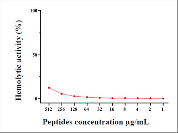
Download figure
Conclusion
Synthetized peptides Neo1, Neo2, Neo3 et Neo4 exhibited inhibitory activities on several strains of bacteria. The use of rich growth media inhibited, directly or indirectly, the antibacterial activity of peptides. We conclude that these peptides can be considered as an antibacterial agent and that, like many antibiotics, they have conditions of activity. These conditions are not met in standard micro dilution assays, but are nonetheless relevant to their natural role and to several therapeutic applications.
References
- 1.Mancuso G, Midiri A, Gerace E, Biondo C. (2021) Bacterial Antibiotic Resistance: The Most Critical Pathogens. Pathogens 10, 1310-10.
- 2.Ho T N T, Turner A, S H Pham, H T Nguyen, Nguyen L T T et al. (2023) Cysteine-rich peptides: From bioactivity to bioinsecticide applications. Toxicon 230, 107173-10.
- 3.Baindara P, Kapoor A, Korpole S, Grover V. (2017) Cysteine-rich low molecular weight antimicrobial peptides from Brevibacillus and related genera for biotechnological applications. World J Microbiol Biotechnol 33, 124-10.
- 4.Takeuchi H. (2021) The role of diverse LURE-type cysteine-rich peptides as signaling molecules in plant reproduction. Peptides (N.Y.) 142, 170572-10.
- 5.Larsson D G J, Flach C-F. (2022) Antibiotic resistance in the environment. Nat Rev Microbiol 20, 257-269.
- 6.D Chinemerem Nwobodo, M C Ugwu, C Oliseloke Anie, Al‐Ouqaili M T S, J Chinedu Ikem et al. Antibiotic resistance: The challenges and some emerging strategies for tackling a global menace. J Clin Lab Anal 36.
- 7.C M Gill, Dolan L, L M Piggott, A M McLaughlin. (2022) New developments in tuberculosis diagnosis and treatment. Breathe 18, 210149-10.
- 8.Wei M, Zhao Yongjie, Qian Zhuoyu, Yang Biao, Xi J et al. (2020) Pneumonia caused by Mycobacterium tuberculosis. Microbes Infect 22, 278-284.
- 10.S Reza Mortazavi-Tabatabaei, Ghaderkhani J, Nazari A, Sayehmiri K, Sayehmiri F et al. (2019) Pattern of antibacterial resistance in urinary tract infections: A systematic review and meta-analysis. Int J Prev Med 10, 169-10.
- 11.Li Y, Kumar S, Zhang L, Wu H. (2022) Klebsiella pneumonia and Its Antibiotic Resistance: A Bibliometric Analysis, Biomed Res Int. 1-10.
- 12.REIS A C C, SANTOS S R da S, SOUZA S C de, M G SALDANHA, T N PI et al. (2016) . CIPROFLOXACIN RESISTANCE PATTERN AMONG BACTERIA ISOLATED FROM PATIENTS WITH COMMUNITY-ACQUIRED URINARY TRACT INFECTION, Rev Inst Med Trop Sao Paulo 58, 10-1590.
- 13.Mintz I, Chowers M, Obolski U. (2023) Prediction of ciprofloxacin resistance in hospitalized patients using machine learning. Communications Medicine 3, 43-10.
- 14.Shariati A, Arshadi M, M A Khosrojerdi, Abedinzadeh M, Ganjalishahi M et al. (2022) The resistance mechanisms of bacteria against ciprofloxacin and new approaches for enhancing the efficacy of this antibiotic. Front Public Health 10, 10-3389.
- 15.Sharma J, Sharma D, Singh A, Sunita K. (2022) Colistin Resistance and Management of Drug Resistant Infections. Canadian Journal of Infectious Diseases and Medical Microbiology 1-10.
- 16.S P Doijad, Gisch N, Frantz R, B V Kumbhar, Falgenhauer J et al. (2023) Resolving colistin resistance and heteroresistance in Enterobacter species. Nat Commun 14, 140-10.
- 18.Aminov R. (2022) Metabolomics in antimicrobial drug discovery. Expert Opin Drug Discov 17, 1047-1059.
- 19.V M Chernov, O A Chernova, A, L, R I Aminov. (2019) Omics of antimicrobials and antimicrobial resistance. Expert Opin Drug Discov 14, 455-468.
- 20.Shen X, Shen J, Pan Y, Cheng J, Chai J et al. (2021) Clinical diagnosis and treatment of common respiratory tract infections in relation to microbiological profiles in rural health facilities in China: implications for antibiotic stewardship. BMC Fam Pract 22, 10-1186.
- 21.Clavenna A, Bonati M. (2011) Differences in antibiotic prescribing in paediatric outpatients. Arch Dis Child 96, 590-595.
- 22.H M Crowe, Quintiliani R. (1995) Antibiotic formulary selection. Medical Clinics of North America 79, 463-476.
- 23.M S Pulia, S C Keller, C J, Jump R L P, T. (2020) Antibiotic Stewardship for Older Adults in Ambulatory Care Settings: Addressing an Unmet Challenge. J Am Geriatr Soc 68, 244-249.
- 24.Walther R, Rautio J, A N Zelikin. (2017) Prodrugs in medicinal chemistry and enzyme prodrug therapies. Adv Drug Deliv Rev 118, 65-77.
- 25.M R Yeaman, N Y Yount. (2003) . Mechanisms of Antimicrobial Peptide Action and Resistance, Pharmacol Rev 55, 27-55.
- 26.Khazaei-poul Y, Farhadi S, Ghani S, S A Ahmadizad, Ranjbari J. (2020) Monocyclic Peptides: Types, Synthesis and Applications. Curr Pharm Biotechnol 22, 123-135.
- 27.Yi J, Kim C, C A Gelfand. (2007) Inhibition of Intrinsic Proteolytic Activities Moderates Preanalytical Variability and Instability of Human Plasma. J Proteome Res 6, 1768-1781.
- 28.Böttger R, Hoffmann R, Knappe D. (2017) Differential stability of therapeutic peptides with different proteolytic cleavage sites in blood, plasma and serum. e0178943. https://doi.org/10.1371/journal.pone.0178943 , PLoS One 12.
- 29.Huan Y, Kong Q, Mou H, Yi H. (2020) Antimicrobial Peptides: Classification, Design, Application and Research Progress in Multiple Fields. https://doi.org/10.3389/fmicb.2020.582779 , Front Microbiol 11.
- 30.Le C-F, Fang C-M, S D. (2017) Intracellular Targeting Mechanisms by Antimicrobial Peptides. Antimicrob Agents Chemother 61, 10-1128.
- 31.Shu G, Chen Y, Liu T, Ren S, Kong Y. (2019) Antimicrobial Peptide Cathelicidin-BF Inhibits Platelet Aggregation by Blocking Protease-Activated Receptor 4. Int J Pept Res Ther 25, 349-358.
- 32.Kuriakose A, Chirmule N, Nair P. (2016) Immunogenicity of Biotherapeutics: Causes and Association with Posttranslational Modifications. J Immunol Res 1-18.
- 33.Groot A S De, B J Roberts, Mattei A, Lelias S, Boyle C et al. (2023) Immunogenicity risk assessment of synthetic peptide drugs and their impurities. Drug Discov Today 28, 103714-10.
- 34.C A Roque-Borda, L P Pereira, Guastalli E A L, N M Soares, Mac-Lean P A B et al. (2021) HPMCP-Coated Microcapsules Containing the Ctx (Ile21)-Ha Antimicrobial Peptide Reduce the Mortality Rate Caused by Resistant Salmonella Enteritidis in Laying Hens. Antibiotics 10, 616-10.
- 35.König W, Geiger R. (1970) Eine neue Methode zur Synthese von Peptiden: Aktivierung der Carboxylgruppe mit Dicyclohexylcarbodiimid unter Zusatz von 1‐Hydroxy‐benzotriazolen. 788–798. https://doi.org/10.1002/cber.19701030319 , Chem Ber 103.
- 36.P C Schreckenberger, M J Binnicker. (2011) Optimizing Antimicrobial Susceptibility Test Reporting. J Clin Microbiol 49, 10-1128.
- 37.G A Pankey, L D Sabath. (2004) Clinical Relevance of Bacteriostatic versus Bactericidal Mechanisms of Action in. the Treatment of Gram‐Positive Bacterial Infections, Clinical Infectious Diseases 38, 864-870.
- 38.Qin N, Tan X, Jiao Y, Liu L, Zhao W et al. (2014) RNA-Seq-based transcriptome analysis of methicillin-resistant Staphylococcus aureus biofilm inhibition by ursolic acid and resveratrol. Sci Rep 4, 5467-10.
- 39.Jia F, Wang J, Peng J, Zhao P, Kong Z et al. (2017) D-amino acid substitution enhances the stability of antimicrobial peptide polybia-CP. 916–925. https://doi.org/10.1093/abbs/gmx091 , Acta Biochim Biophys Sin (Shanghai) 49.
- 40.Deracinois B, Matéos A, Romelard A, Boulier A, Auger J et al. (2021) Partial-, Double-Enzymatic Dephosphorylation and EndoGluC Hydrolysis as an Original Approach to Enhancing Identification of Casein Phosphopeptides (CPPs) by Mass Spectrometry. Foods 10, 2134-10.
- 41.Torrent M, V M Nogués, Boix E. (2009) A theoretical approach to spot active regions in antimicrobial proteins. BMC Bioinformatics 10, 373-10.
- 42.Gautier R, Douguet D, Antonny B, Drin G. (2008) HELIQUEST: a web server to screen sequences with specific α-helical properties. Bioinformatics 24, 2101-2102.
- 43.Vallelian F, P W Buehler, D J Schaer. (2022) Hemolysis, free hemoglobin toxicity, and scavenger protein therapeutics. Blood 140, 1837-1844.