
Ultrasound Guided Pericardial Effusion Training Model for Neonates
Abstract
This methods article presents a neonatal pericardial effusion training model using ultrasound. It details materials, construction, and training objectives to support procedural competence.
Author Contributions
Copyright © 2024 Monalisa Patel
 This is an open-access article distributed under the terms of the Creative Commons Attribution License, which permits unrestricted use, distribution, and reproduction in any medium, provided the original author and source are credited.
Competing interests
The authors declare no conflict of interest.
Citation:
Background
Cardiac tamponade due to pericardial effusion is a life-threatening emergency in which pericardiocentesis may be required. 1
It can cause hemodynamic collapse without immediate intervention. Although the incidence is low1 in comparison to other life-threatening medical conditions encountered in the neonatal intensive care unit, neonatologists must be familiar and comfortable with the management of significant pericardial effusion causing tamponade physiology2. Since it is encountered infrequently, many providers may have little or no experience in managing the condition and performing a lifesaving pericardiocentesis.
Pericardiocentesis has been historically performed as a “blind” procedure, however the availability of bedside ultrasound has radically increased the success rates of the procedure2. As ultrasound guidance lowers the complication rates and increases the patient’s safety, pericardiocentesis should be performed under ultrasound guidance. Neonatologists actually need to train in emergency pericardiocentesis.3 The use of ultrasound guidance may also allow for more real-time feedback for the trainees. There are various simulation models for pericardial effusion drainage available, for training under ultrasound guidance, however majority of them are geared towards larger children/adults. There is a need for creating a ultrasound guided neonatal pericardiocentesis model that is reusable and cost effective.
Objective
There is a need for the development of a low cost, durable, and high-fidelity, procedure-specific simulation models in neonatology. 4 I wanted to create such pericardial effusion model to train neonatologists and neonatal trainees.
My objective was to create a re-usable unperishable model for ultrasound guided pericardiocentesis. I performed a literature review on various existing pericardiocentesis models.
Models described in the literature have used items such as pork ribs, tofu, gelatin, ballistic gel or gel wax to serve as the thoracic cavity medium and items such as golf balls, balloons, saline bags, or ping pong balls for ventricle or pericardial effusion . It appeared that the model created by Daly et al would be feasible to tailor for our neonatal specific pericardial effusion simulation model.
Young et al had devised a modification of Daly’s model that offers the benefit of more realistic external landmarks. I also adapted some of the suggestions as made by Young et al 5
Materials
| Medical Gel | Humimic medical | 117$ |
| Flesh Tone dye | Humimic medical | 35$ |
| Halloween skeleton | Amazon | 25$ |
| Saline bags | IV supply clinic | 30$ |
| Three way stop cock | Amazon | 10$ |
| Ping Pong balls | Amazon | 13$ |
| Food coloring | Walmart | 5$ |
| Needle 18G | Amazon | 12$ |
| Cyanoacrylate superglue | Amazon | 14$ |
| Jorvet universal IV set | Amazon | 5$ |
Creation of the model
A lot of the steps are very similar to Daly Et Al’s pericardiocentesis model however the size of the rib cage, the container, the saline bags were all tailored specific to a neonatal model for pericardiocentesis.1
Simulate the neonatal thorax: The Halloween skeleton with a rib cage transverse diameter 14 cm was dismantled manually from its connected arms and posterior ribs. Such that only the sternum and anterior ribs were left to create the model. The rib cage is placed in a rectangular glass baking tray that is 20x15x7 cm (LxWxH) sternum down
Simulate the ventricle: make two small holes in a ping pong ball using 18G needle. Fill the red colored dyed water in one hole using the needle and a syringe. The other hole will allow the air from the ping pong ball to escape. Once nearly all air is cleared, and the ball filled, sealed the holes using cyanoacrylate glue.
Simulate the pericardial effusion: 50ml saline bag was used to mimic effusion. The bag was spiked with universal IV set. The other end of the IV set was attached to a 3-way stop cock and flush the tubing of air. The three way stop cock was then connected to a 50ml syringe. Manipulate the saline bag when flushing the line to ensure that as much air is removed from the bag as possible. 1 The top part of the spike was covered with yellow colored dye before spiking the saline bag, which then got dissolved in the saline bag giving it the straw color of effusion. The 50 ml syringe is filled with yellow colored water and the 3-way stop cock left “off “to the model. This syringe will serve in the future to refill the saline bag with more yellow colored water/effusion as practitioners start draining the pericardial fluid during their practice sessions. Below are the exact steps I followed in creating the gel model of neonatal thorax with pericardial effusion.
1. Simulate the skin/ subcutaneous tissue using medical gel: I placed the anterior rib cage in the glass container, with sternum down. I put that set up aside. I used total 3 packs of humimic medical gel. I placed all three packs of the gel in a large steel pot on a stove top. I used kitchen thermometer to measure the temperature of gel. Once it reached 250F I added skin tone dye in it and mixed it thoroughly while making sure there were no bubbles. I poured a third amount of gel in the glass baking container with the rib cage in it. I left it in a cool environment until the gel has become firm and taken the form of the container. This took about 45-60 minutes. Picture 1
2. Simulate the right ventricle and the effusion: Then I placed the saline bag on the gel in its anatomically correct position of fluid around simulated right ventricle. Then I placed a thin layer of gel on top of saline bag. Once that layer was cool and settled, I placed the ping pong ball on top of the layer covered saline bag. This thin 1-2 cm layer serves as a delineation between the saline bag and the fluid filled ping pong ball. Under ultrasound it may serve as a proxy for the ventricular wall between the ping pong ball/ “ the right ventricle” and the saline bag/ “the effusion”. It is important to remember to place the ball and the bag in as anatomically correct as possible locations of the rib cage Picture 2
3. Simulate the posterior part of rib cage and completing the model: The second pour of the medical gel is placed on top of the ping pong ball and saline bag. Great care is needed at this step to avoid the ball/saline bag from moving away due to hot gel. Hence, they were kept in place using a wooden spatula. At this pour only half of the ping pong ball is intended to be covered with gel. Picture 3
4. The back of rib cage: Once the second pour is getting firm and settled a last final pour is placed to ensure the entire ping pong ball and all the ribs are completely covered by the gel. Picture 4
The pictures of the final version of the model along with the ultrasound images are at the bottom of this article.
Picture 1.The red arrow indicates the right ventricle and the green arrow indicates the pericardial effusion note that the fusion does not appear circumferential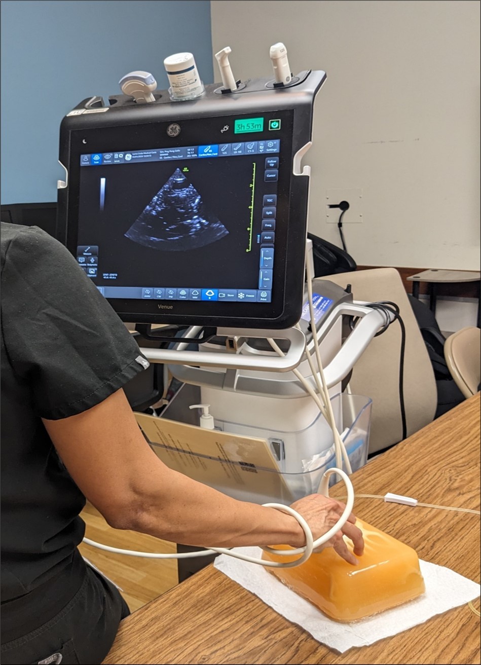
Picture 2.Showing the complete model of the neonatal pericardial effusion. We can see the spiked portion of the sailing bag coming out from the model. We can also see some yellow-colored fluid coming out in the tubing which represents the simulated pericardial effusion fluid. The red arrow shows a three-way stopcock that connects the tubing to a 50 ML syringe. This syringe will be used to refill the saline bag simulating the pericardial effusion once the fluid is decreasing after it's drained by the trainee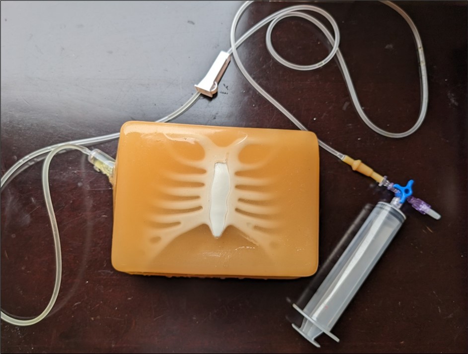
Picture 3.Showing the pericardial effusion from the subcostal view under ultrasound guidance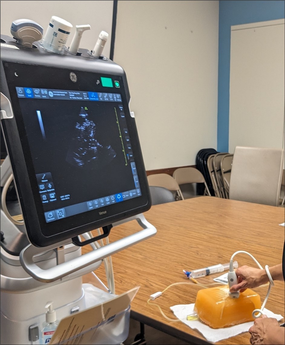
Picture 4.Close view of the height and width of the neonatal pericardial effusion model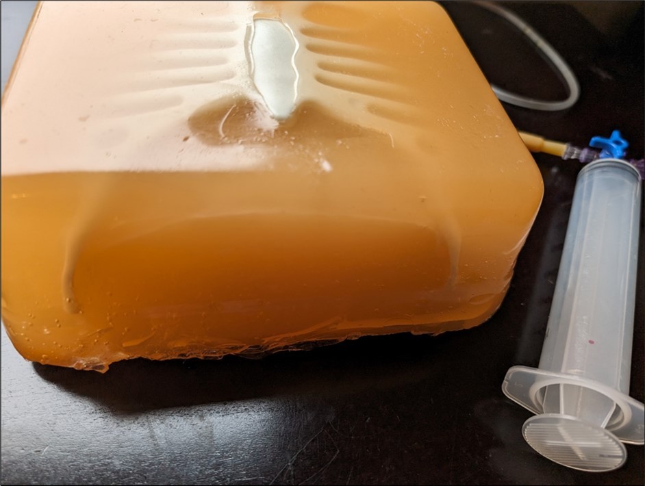
Our experience
Our neonatal gel wax model was used to train fellows and nurse practitioners. A total of 12 fellows, 3 nurse practitioners and 4 attending physicians performed ultrasound guided pericardiocentesis at least twice with successful pericardial fluid tap. These trainees were not exposed to this gel wax model before however they were able to identify the ventricle and presence of fluid around it on ultrasound. We did not perform a statistical analysis due to the limited number of individuals. The gel wax model with saline bag was able to withstand the repeated needle punctures. If during training someone accidentally needle punctures the “ventricle”/the ping pong ball, then they will aspirate red fluid. If someone needle punctures the pericardial fluid/saline bag they will aspirate the yellow fluid. The quality of the ultrasound image did deteriorate though after about 30 punctures. The gel wax though continues to retain its structure with its inbuilt rib cage and saline bag after 4 months of its original creation. It appeared that after the first 30 punctures, the fluid from saline bag started to accumulate in the chest cavity and leak some from the chest wall which may have contributed to the decrease in image quality after those punctures.
Feedback for future models
One of the limitations in this model was that after several needle punctures, the fluid for the effusion was collecting inside the model, and hence interfering with the ultrasound images. So there needs to be a way to create the model such that the fluid has an outlet to drain outside the model. The pericardial effusion that was stimulated by the saline bag appeared more rectangular or longitudinal on the imaging whereas generally the pericardial effusion in the clinical scenario would appear more so circumferential or circular on imaging so we would need to create the model such that either the saline bag is made circular within the model or is replaced by something that makes the fluid/effusion appear circumferential or circular around the simulated ventricle
References
- 1.Daly R, J H Planas, M A Edens. (2017) Adapting Gel Wax into an Ultrasound-Guided Pericardiocentesis Model at Low Cost. , West. J. Emerg. Med 18, 114-116.
- 2.Pezeshki B, Boodaie B, Kessel M, Grewal E, Alexander D. (2021) A Novel, Reusable, Ultrasound-Guided Pericardiocentesis Simulation Model. , J. Ultrasound Med 40, 1657-1663.
- 3.dell’Orto Campo, M. (2013) Assessment of a Low-Cost Ultrasound Pericardiocentesis Model. , Emerg. Med. Int 2013, 376415.