
Abstract
Objectives
The number of cases of cyanoacrylate closure (CAC) system for varicose veins has been increasing worldwide. However, as this is a new treatment method, the potential adverse effects and other details remain unclear. In particular, the cause of inflammation in embolized veins is still under debate.
Methods
We performed a drug-induced lymphocyte stimulation test (DLST) on a patient with allergic-like symptoms after CAC.
Results
The DLST was strongly positive in this case, and the patient underwent total removal of the CAC-filled vein due to difficulty controlling the symptoms with medication. After that the state was recovered and no medication was continued.
Conclusion
We encountered a case that a delayed allergy by CA after CAC treatment developed in, eventually leading to the total removal of the CA-filled vein. It was suggested that with doubting allergic-like symptom after CAC, DLST for CA could show not only the diagnosis of the delayed allergy to CA, but also the later treatment policy with stimulation index (S.I.) in the positive cases.
Author Contributions
Academic Editor: Anubha Bajaj, India.
Checked for plagiarism: Yes
Review by: Single-blind
Copyright © 2023 Hiroyuki Suzuki, et al.
 This is an open-access article distributed under the terms of the Creative Commons Attribution License, which permits unrestricted use, distribution, and reproduction in any medium, provided the original author and source are credited.
Competing interests
The authors have declared that no competing interests exist.
Citation:
Introduction
Varicose veins have established treatment methods, ranging from stripping surgery to radiofrequency and laser ablation. In Japan, these procedures are approved as covered by insurance and are already widely known. And cyanoacrylate endovascular embolization (CAC) has been covered by health insurance since December 2019 and has been available at our hospital since March 2020.
The VenaSealTM Closure System is a treatment in which cyanoacrylate (CA) is injected into the treated vessel to occlude it, thereby eliminating venous regurgitation. A high occlusion rate has been reported.
However, as this is a new treatment method, there is still no clear consensus on its complications and preventive measures.
In the present study, we performed a drug-induced lymphocyte stimulation test (DLST) on a patient with allergic-like symptoms after CAC. The result was positive in this case, and the patient underwent total removal of the CAC-filled vein due to difficulty controlling the symptoms with medication.
About this case, the report was published with the Japanese Journal of Vascular Surgery from our hospital in 2022.
The number of CAC case increases worldwide, meanwhile the clear definition and diagnosis method about, it’s complication, phlebitis anddelayed allergy for CA which is extremely rare but the risk of aggravation, are still controversial.
The case report which shows that DLST was useful for the decision of the diagnosis and of the later treatment policy with this issue, has an important meaning. Such a report was not found globally, therefore we edited it in English and reported it again, adding the next progress of the patient and consideration of the CAC cases with DLST at our hospital, with agreement and cooperation from Nakayama who was a former article lead author.
Case report
Patient:
79 years old, female
Chief complaint:
Redness, swelling, and tenderness on the medial side of the affected (left) thigh after CAC surgery.
History:
None of note, no history of allergies, including drugs.
Current medical history
The patient had varicose veins for 30 years and was recently diagnosed with swelling, fatigue, and limpness in the lower extremities. The patient's initial ultrasonography showed reflux in the left great saphenous vein, and CAC treatment was selected based on the patient's desire for treatment.
On admission, the patient was 155 cm tall, 55 kg, body mass index (BMI) 22.8, blood pressure 130/70 mmHg, pulse 62/min, body temperature 36.5 ℃, and lower limb: varicose vein formation on the inner side of the left lower leg. Swelling of the entire left lower extremity and dilation of superficial veins were noted. The skin on the left lower leg was generally dry.
CEAP classification: C1,2,3, Ep, As, Pr, GSVa, GSVb
Preoperative ultrasonography
The diameter of the left great saphenous vein was 3.5 mm above the thigh, 4.2 mm above the knee, and 2.6 mm at the ankle joint in the upright position; leg milking showed reflux waves from the saphenofemoral venous junction (SFJ) to below the knee. Reflux was not observed in the ankle joint.
Surgical findings
Without premedication, the surgery was initiated in the supine position under local anesthesia and with intravenous anesthesia. The left great saphenous vein was punctured under an ultrasonography approximately 5 cm below the knee. A sheath catheter was inserted into the great saphenous vein toward the central side. The catheter tip was positioned approximately 5 cm peripherally from the SFJ by body surface ultrasonography, and CA filling was started.
After CA filling, the sheath was moved a few centimeters toward the central area and the catheter tip was retracted into the sheath. The sheath and catheter were removed so that no CA remained subcutaneously.
Operation time:
0 hours 36 minutes
CA injection volume:
1.5 mL, CAC treatment
length:
40 cm
Postoperative course
Immediately after surgery, the patient was managed without wearing elastic stockings.
At the outpatient visit on the 4th postoperative day, postoperative ultrasonography confirmed the disappearance of the left great saphenous vein reflux. There were no subjective symptoms, such as redness or pain in the left medial thigh. Blood analysis showed a white blood cell (WBC) count of 5200/μL (neutrophils, 62%; lymphocytes, 31%; eosinophils, 3%), which was within the normal range, and no noticeable increase in the C-reactive protein (CRP) level was observed.
However, erythema appeared near the left knee joint at night on the same day. Gradually, erythema expanded to the entire medial thigh along the great saphenous vein on the affected side.
On the seventh postoperative day, redness, pain, and a mild burning sensation appeared on the medial side of the left thigh. The symptoms were most severe near the knee joint. An allergic reaction by CA was doubted, therefore DLST for CA was performed.
The patient was started on the antihistamine fexofenadine hydrochloride (120 mg/day) and a cetaminophen (600 mg/day). Symptoms began to improve four days later, but the medication was continued.
The symptoms had disappeared at the time of the outpatient visit on the 17th postoperative day. It was therefore decided to discontinue the oral administration of the medication. When she bathed in the evening of the same day, an intensely itchy wheal rash appeared on her entire body, including her face.
The rash did not improve 21 days after surgery (Figure 1), so the patient was started on cerestamine-containing steroids (3 tablets/day). Three days after the start of the medication, the rash symptoms decreased; however, the patient continued to suffer from a severely pruritic rash that easily recurred throughout her body.
Figure 1.Swelling of body, 21 days after CAC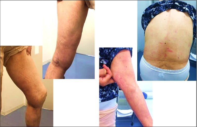
Around that time, the DLST results revealed that the stimulation index (S.I.) was 690%, a strong positive result. The patient was referred to the Department of Allergy and Allergology at our hospital, and olopatadine hydrochloride (10 mg/day), an antihistamine and antiplatelet activating factor was added to her medication.
Since the patient's generalized rash did not improve, it was decided to remove the full-length CA-filled vein.
On the 45th postoperative day, the left CA-filled great saphenous vein was removed under general anesthesia. A segmental incision was made at the perforated branch site. The vein was removed several centimeters from the CA-filled site using a vein harvester. All the CA filling sites were confirmed in the vein that was removed. The pathology results of the extracted vein showed lymphocytic infiltration, histiocyte reaction, and phagocytosis of the foreign body (Figure 2). However, this finding alone does not rule out allergy.
Figure 2.Pathological results of the vein: severe inflammatory change with granuloma formation of the vein
After the surgery, a mild rash appeared on the buttocks and sacral region but improved with olopatadine hydrochloride (10 mg/day) and prednisolone (started at 15 mg/day). The steroid medication was gradually tapered off after ten days. Olopatadine hydrochloride was discontinued after confirming that the patient's condition remained stable after being allowed to bathe. The patient had been taking this drug for 50 days.
Since then, the patient's condition has remained stable without recurrence.
From the first day of CAC, we confirmed that the patient's condition had stabilized after removing the drug, and we checked blood samples as necessary. At that time, no abnormally high WBC or CRP levels were confirmed, and the neutrophil/eosinophil ratio in the WBC fraction remained normal.
The S.I. of the DLST results decreased after the removal of the CA-filled vein and eventually became negative, as follows: 690% after CAC surgery, 260% six months after the removal of the CA-filled vein, and 175% (normal value) one year after removal of the CA-filled vein.
Discussion
CAC treatment eliminates the reflux of superficial veins that cause varicose veins by filling targeted veins with CA. CA is a strong, fast-curing adhesive used in industrial, medical, and household applications. It is often used as a nail adhesive in the beauty industry, including nail art.
Various reports of allergic reactions after CAC are recognized.
Morrison et al. found no allergic reactions to the skin or deep tissue in 108 CAC cases.1
On the other hand, there are reports of hypersensitivity reactions to subcutaneous tissue at the time of delivery of the CAC system2 and reports of generalized urticaria one week after CAC3. Park et al. reported an unusual phlebitis-like reaction that occurred in 25% of CAC cases, in which they postulated a type IV hypersensitivity reaction (delayed allergy) as the mechanism4. The above complication is also mentioned in the "Guidelines for Endovascular Therapy with Cyanoacrylate Adhesives for Varicose Veins" in Japan and is called phlebitis, the mechanism of which is still unclear5.
In our case, erythema and swelling appeared on the medial side of the affected thigh seven days after surgery, leading us to suspect a type IV hypersensitivity reaction. A DLST was performed using the CA used in the treatment to clarify this. This test is useful to determine whether a particular drug is involved in drug-induced allergic symptoms, especially liver damage and hematopoietic disorders caused by type IV allergic mechanisms. The sensitivity and specificity of the test are 60–70% and 85%, respectively, depending on the target drug6.
The DLST is more likely to be negative when steroids, antitumor agents, or immunosuppressive agents are used, whereas it may be positive when non-steroidal anti-inflammatory drugs are used. In our case, the patient took antihistamines but no other anti-inflammatory drugs.
As the patient was not taking any medications that could have affected the results when the test was performed, the results were considered highly reliable. Therefore, a positive DLST means indicated a delayed allergy for CA.
Jones et al. stated that they administered oral steroids and antihistamines when a post-CAC hypersensitivity reaction was suspected7. Our patient was treated with a combination of oral antihistamines and steroids and the skin rash disappeared once, but recurred as a generalized rash, including on the face. Furthermore, the rash was a highly pruritic wheal, without erythema, swelling, or pain on the medial side of the affected thigh seen in the acute phase of phlebitis. Jones et al. stated that the incidence of hypersensitivity reactions leading to removing the treated vein is less than 1 in 10,000, according to the manufacturer's publication. Therefore, this case was regarded as such an incident.
The total number of CAC cases at our hospital in the end of December, 2022, was 368. Meanwhile 27 cases doubted an allergic reaction, including extremely slight symptoms, were detected (7.3% during all cases), and all of them were performed DLST for CA. As a result, 3 cases (0.8% during all cases) were detected as positive (more than 180% of S.I. ).
In each DLST-negative cases, redness, swelling and pain on the medial side of the affected thigh occurred 6-8 days after CAC, and the symptoms were improved by the internal use of antihistamines and non-steroidal anti-inflammatory drugs. These cases are considered as phlebitis.
As a breakdown of 3 DLST-positive cases, one case was strong positive S.I. 690%, and the other two cases were S.I. 288% and 237%. Not only a redness, swelling and pain on the medial side of the affected thigh, but also the wheal with the itch were detected with all of these 3 cases. The systemic wheal was found on only the DLST strong positive case. About the DLST slight positive cases, the relief of symptom was detected by the internal use of antihistamines and non-steroidal anti-inflammatory drugs. On the other hand, about the DLST strong positive case, the improvement and stabilization of the symptom were not detected, even if steroids were given. The disease severity varies according to each S.I. value in the DLST-positive case, that mean delayed allergy for CA. The removal of the CA-filled great saphenous vein could be necessary in the case of the abnormal high S.I. value like this case, without the improvement of the symptom only by medication treatment. Thus, it was suggested that DLST could show not only the diagnosis of the delayed allergy, but also the later treatment policy with S.I. value in the positive cases.
In addition, our patient's DLST SI value gradually decreased after removing the CA-filled vein and eventually became negative. This is consistent with the patient's skin symptoms, which also lend credence to the DLST.
Although not a frequent occurrence, it would be ideal to confirm the presence of an allergic reaction to CA before surgery to avoid hypersensitivity reactions after CAC. However, Jones et al. stated no valid evidence exists for a CA patch test routinely performed before CAC surgery7. For this reason, our clinic now asks patients to indicate on the initial medical questionnaire whether or not they have a history of allergy and the nature of their symptoms.
Given the current difficulty in confirming the presence or absence of preoperative hypersensitivity reactions to CA, the DLST for CA may be useful in determining the course of treatment in the event of a postoperative allergic-like reaction.
Conclusion
We encountered a case that a delayed allergy by CA after CAC treatment developed in, eventually leading to the total removal of the CA-filled vein.
It was suggested that with doubting allergic reaction-like symptom after CAC, DLST for CA could show not only the diagnosis of the delayed allergy to CA, but also the later treatment policy with S.I. value in the positive cases.
The accumulation of the further cases is necessary to establish the clear S.I. value of DLST for CA, for judging of the disease severity.
References
- 1.Morrison N, Gibson K, McEnroe. (2015) Randomized trial comparing cyanoacrylate embolization and radiofrequency ablation for incompetent great saphenous veins(Veclose). J Vasc Surg. 61, 985-94.
- 3.Gibson K, Ferris B. (2017) Cyanoacrylate closure of incompetent great, small and accessory saphenous veins without the use of post-procedure compaserrion: Initial outcomes of a post-market evaluation of the VenaSeal System (the WAVES Study). Vascular. 25, 149-56.
- 4.Park I, Jeong M H, Park C J. (2019) Clinical Features and Management of “Phlebitis-like abnormal reaction” after cyanoacrylate closure for the treatment of incompetent saphenous veins. Ann Vasc Surg. 55, 239-45.
- 5.Hirokawa M, Satogawa H, Yasugi T. (2020) Cyanoacrylate Glue Closure for varicose Veins:. , Concensus Guidelines of the Japanese Society of Phlebology. Jpn J Phlebol 31, 141-52.